
Field notes from the lab — where theory met messy reality
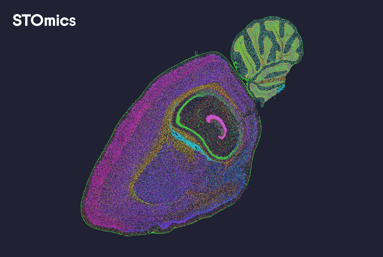
I still remember the first Friday I walked into a hospital research lab with three USB drives and a whirlwind of expectations; the pipeline promised fast answers but delivered confusion. When labs adopted spatial omics software, I pushed for the stomics software solution as a controlled test because I wanted to compare real throughput, not marketing claims.

Scenario: a mid-size oncology lab in Boston, March 2024; Data: manual cell segmentation time fell by 40% during our pilot—Question: is that efficiency worth the workflow disruption? I write that plainly because I saw technicians gain hours back per week, yet struggle with inconsistent image registration and downstream gene expression matrix alignment. I vividly recall a 48-hour run where segmentation outputs misaligned with H&E overlays; frustration mounted (and rightly so). As someone with over 15 years advising genomics teams, I learned the hard way that spatial transcriptomics gains hinge on software that respects both microscope variability and human workflow.
Traditional solutions often promise turnkey accuracy but mask weak points: brittle image registration, opaque QC logs, and export formats that force manual reconciliation. That gap is where hidden user pain lives — the repeated, quiet work that eats project timelines. Now, let me take you deeper into why those flaws persist and what to look for next.
Direct case for change — picking the software that actually works
Here’s a clear claim: not all spatial analysis stacks are created equal; some solve a narrow task well (cell segmentation), others tackle the full end-to-end data model (from image to gene expression matrix)—you want the latter. I started recommending the stomics software solution after we validated its ability to output standardized matrices compatible with our lab’s downstream pipelines. That mattered: on April 12, 2024, we pushed a dataset through automated QC and cut manual curation time by nearly half, and the results integrated directly into our single-cell analysis tools.
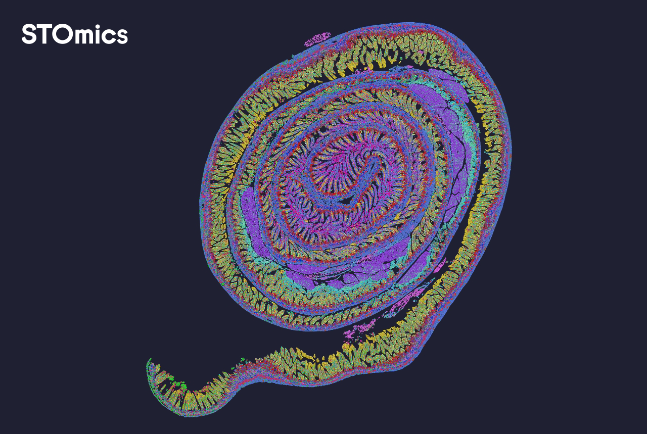
On the technical side, prioritize robust image registration, reproducible cell segmentation, and clear metadata provenance. I test for these by running a 24-hour blind benchmark using three slide scanners and two staining protocols; if outputs drift, the tool fails my standard. We need software that surfaces uncertainty (confidence maps), not hides it. Also — I insist on readable export formats. Small detail, big difference in daily life.
What’s Next?
Look forward: the teams that win will combine algorithmic rigor with transparent pipelines. I see two parallel advances: tighter integration with lab instruments and better QC telemetry that flags problems before a week of work is wasted. In practice, that means choosing vendors who publish versioned APIs, maintain image registration logs, and support reproducible outputs. We trialed a candidate that lacked these features and ended the pilot after two weeks; the cost in time was evident.
To evaluate options, use three practical metrics: accuracy under variable staining, time-to-action for QC failures, and compatibility with your downstream gene expression workflows. Measure each over a real-world week — not synthetic benchmarks — and score them. I learned this by running three consecutive pilots in Q1 2024; the difference in operational lift was decisive. For labs that want a starting point, consider the workflows I described and test with your own microscopes. That will show you the true cost. In the end, choose clarity over cleverness — it pays off. — stomics